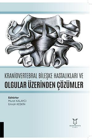
Kraniovservikal Bileşke Embriyolojisi
Synopsis
Authored by İskender Samet DALTABAN and Mehmet Selim GEL, this work presents a comprehensive literature review on the embryological development, anatomy, and biomechanics of the craniovertebral junction (CVJ), a complex structure of vital importance to neurosurgical clinicians due to the critical neural and vascular elements it surrounds. Emphasizing that understanding developmental stages is essential for grasping congenital and acquired anomalies, the text detailedly addresses the presomitic stage beginning with gastrulation, primary segmentation shaped by somitogenesis, and mesenchymal differentiation. The chapter specifically examines the unique developmental features of the CVJ derived from four occipital and the first three cervical somites, highlighting the key role of the proatlas sclerotome in the formation of the basiocciput, apical segment of the dens, and occipital condyles. Furthermore, it demonstrates the contributions of the first and second cervical sclerotomes to the development of vertebral bodies, posterior arches, the dens structure, as well as the alar and transverse ligaments. Drawing attention to the clinical implications of disruptions during developmental stages, this resource serves as an essential reference for neurosurgery and anatomy literature.
References
Dietrich S, Kessel M. Thevertebralcolumn. In: Thorogood P, editor. Embryos, genesandbirthdefects. Chichester: Wiley; 1997. pp. 281–302.
Pang D, Thompson DN. Embryology and bony malformations of the craniovertebral junction. Childs Nerv Syst. 2011;27(4):523–564. doi: 10.1007/s00381-010-1358-9.
Davis GK, Patel NH. Theoriginandevolution of segmentation. Trends Cell Biol. 1999;24(12):M68–72. doi: 10.1016/S0962-8924(99)01663-3.
Keynes RJ, Stern CD. Mechanisms of vertebratesegmentation. Development. 1988;103:413–429.
Bellairs R. Thesegmentation of somites in thechickembryo. Bolletinodizoologia. 1980;47:245–252.
Huang R, Zhi Q, Ordahl CP, Thefate of thefirstaviansomite. AnatEmbryol (Berlin) 1997;195:435–449. doi: 10.1007/s004290050063.
Christ B, Wilting J. Fromsomitestovertebralcolumn. Ann Anat. 1992;174:23–32.
Christ B, Jacob HJ, Jacob M. On theformation of themyotomes in avianembryos. An experimentalandscanningelectronmicroscopestudy. Experentia. 1978;34:514–516. doi: 10.1007/BF01935963.
Ebner E. Urwirbel und Neugliederung der Wirbelsäule. Sitzungsber Akad Wiss Wein III. 1888;97:194–206.
Müller F, O’Rahilly R. Segmentation in staged human embryos: the occipitocervical region revisited. J Anat. 2003;203:251–258. doi: 10.1046/j.1469-7580.2003.00219.
Ganguly DN, Roy KKS: A study on thecraniovertebraljoint in man. AnatAnzBd 114:433–452, 1964
Muller F, O'Rahilly: Thehumanchondrocranium at theend of the embryonic period proper with particular reference to the nervoussystem. Am J Anat 159:33–58, 1980.
Melsen B: Thecranialbase. ActaOdontScand 32:1–126, 1974.
Gladstone J, Erickson-Powell W: Manifestation of occipitalvertebraandfusion of atlas withoccipital bone. J Anat&Physiol 49:190–199, 1914–1915
Wilting J, Ebensperger C, Müller TS. Pax-1 in the development of the cervico-occipital transitional zone. Anat Embryol (Berl) 1995;192:221–227. doi: 10.1007/BF00184746
Menezes AH. Craniocervical developmental anatomy and its implications. Childs Nerv Syst. 2008;24(10):1109–1122. doi: 10.1007/s00381-008-0600-1.
Curtis Edward Offiah, Emily Day The craniocervical junction: embryology, anatomy, biomechanics and imaging in blunt trauma Insights Imaging. 2017 Feb; 8(1): 29–47. Published online 2016 Nov 4. doi: 10.1007/s13244-016-0530-5