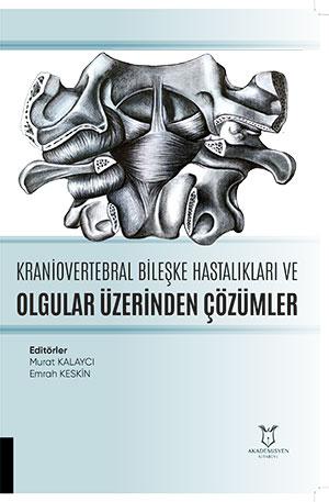
Kraniovertebral Bileşke Anatomisi
Synopsis
The chapter titled "Craniovertebral Junction Anatomy" authored by Dr. Emrah Keskin details this complex and vital transition region where the skull, upper cervical spine, brain, and spinal cord meet in all its anatomical aspects. Housing the spinal cord and cranial nerves as well as critical vascular structures that feed the brain and cervical spinal cord parenchyma, the craniovertebral junction (CVJ) carries significant potential for morbidity and mortality due to its structural complexity. The work thoroughly examines the bone anatomy of the CVJ, including the squamous, basal, and condylar parts of the foramen magnum, its significance in posterior and transoral surgical approaches, the morphological features of the atlas (C1) and axis (C2) vertebrae, and clinical variations such as the Kimmerle anomaly. In addition, the anatomical course along the sulcus arteriosus and foramen transversarium as well as the branches of the vertebral artery, which originates from the subclavian artery and divides into four distinct segments (V1-V4), are explained. The atlanto-occipital, median atlanto-axial, and lateral atlanto-axial joints, key ligaments maintaining stability such as the alar ligament and cruciforme ligament of the atlas, along with the M. longus colli, M. rectus capitis, and suboccipital muscle groups are comprehensively covered. Offering complete foundational anatomical knowledge essential for determining etiology and selecting appropriate surgical treatment in CVJ pathologies, this study serves as an essential reference source in the field of neurosurgery.
References
Menezes AH, Traynelis VC. Anatomy and biomechanics of normal craniovertebral junction (a) and biomechanics of stabilization (b). Childs Nerv Syst. 2008 Oct;24(10):1091-100. doi: 10.1007/s00381-008-0606-8. Epub 2008 Apr 4. PMID: 18389261.
Brasch JC (1958) Cunningham’s manual of practical anatomy. Oxford University Press, London, pp 258–295
VanGilder JC, Menezes AH, Dolan K (1987) The craniovertebral junction and its abnormalities. Futura, New York, pp 1–255
Salas E, Sekhar LN, Ziyal IM, et al. Variations of the extreme-lateral craniocervical approach: anatomical study and clinical analysis of 69 patients. J Neurosurg. 1999 Apr;90(2 Suppl):206-19. doi: 10.3171/spi.1999.90.2.0206. PMID: 10199250.
Schmidek HH. (2000) Operative Neurosurgical Tecniques. Indications, Methods and Results. Fourth Edition, Volume 2. W.B. Saunders Company. Philadelphia. s:1923-33
Türe U, Pamir MN. Extreme Lateral-Transatlas Approach for Resection of the Dens of the Axis. J Neurosurg. 2002 Jan;96(1 Suppl):73-82
Wysocki J, Bubrowski M, Reymond J, et al. Anatomical variants of the cervical vertebrae and the first thoracic vertebra in man. Folia Morphol (Warsz). 2003 Nov;62(4):357-63. PMID: 14655117.
Chitroda PK, Katti G, Baba IA, et al. Ponticulus posticus on the posterior arch of atlas, prevalence analysis in symptomatic and asymptomatic patients of gulbarga population. J Clin Diagn Res. 2013;7(12):3044-3047. doi:10.7860/JCDR/2013/6795.3847
Kuhta P, Hart J, Greene-Orndorff L, et al. The prevalence of posticus ponticus: retrospective analysis of radiographs from a chiropractic health center. J Chiropr Med. 2010;9(4):162-165. doi:10.1016/j.jcm.2010.07.004
Taitz C, Nathan H. Some observations on the posterior and lateral bridge of the atlas. Acta Anat (Basel). 1986;127(3):212-217. doi:10.1159/000146284
Bayrakdar IS, Miloglu O, Altun O, et al. Cone beam computed tomography imaging of ponticulus posticus: prevalence, characteristics, and a review of the literature. Oral Surg Oral Med Oral Pathol Oral Radiol. 2014;118(6):e210-e219. doi:10.1016/j.oooo.2014.09.014
Cho YJ. Radiological analysis of ponticulus posticus in Koreans. Yonsei Med J. 2009;50(1):45-49. doi:10.3349/ymj.2009.50.1.45
Gürçay, A.G., Okutan, M.Ö. Kraniyovertebral Bileşkenin Cerrahi Anatomisi, 2015;12(2)
Arıncı K, Elhan A. (2014) Anatomi. Ankara: Güneş Kitabevi: 60
Lasjaunias P, Berenstein A. Surgical Neuro-angiography Volume 3: Functional Vascular Anatomy of Brain, Spinal Cord and Spine. Berlin Heidelberg: Springer-Verlag;1990;1-38, 199-222
Biçeroğlu H, Hancı M: Omurganın Cerrahi Anatomisi. İntertıp Yayınevi, 2014: 69-73