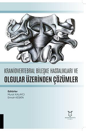
Kranioservikal Bileşke Anomalileri ve Olgular Üzerinden Çözümler (Endoskopik Yaklaşım)
Synopsis
The craniovertebral junction (CVJ) is a complex and critical anatomical region extending from the occipital bone to the C2-C3 intervertebral disc level, containing vital neurovascular structures. Congenital, inflammatory, neoplastic, or traumatic pathologies arising in this area can compress the cervicomedullary junction and lower cranial nerves, leading to severe clinical manifestations such as myelopathy, cranial neuropathies, vertebrobasilar insufficiency, and even sudden death. Although the traditional transoral microsurgical approach was considered the gold standard for many years, it carries significant disadvantages, including oral flora contamination, elevated risk of CSF leaks and meningitis, prolonged intubation, and tissue edema. In contrast, the pure endoscopic endonasal approach—which has gained widespread adoption in recent years—utilizes natural anatomical corridors without requiring skin incisions, offering enhanced illumination and a broad panoramic view of deeply located lesions. By avoiding the oral cavity, this technique eliminates the risks of swallowing and breathing difficulties, thereby enabling early oral intake, faster recovery, and shorter hospital stays. In preoperative planning, radiological lines and angles—such as the palatine line, nasopalatine line, naso-axial line, and Wackenheim angle—are meticulously evaluated to determine the exact inferior and superior boundaries of the lesion. Furthermore, preserving the anterior arch of the atlas during endoscopic endonasal surgery prevents craniocervical instability, thereby eliminating the need for additional posterior fusion.1
References
Dagtekin A, Avcı E, Kara E, et al. Posterior Cranial Fossa Morphometry in Symptomatic Adult Chiari I Malformation Patients: Comperative Clinical and Anatomical Study. Clinical Neurology and Neurosurgery. 2011; 113(5):399-403.
Şengül G, Kadıoğlu HH. Morphometric Anatomy of the Atlas and Axis Vertebrae. Turkish Neurosurgery. 2006; 16(2):69-76.
Tanrıverdi O, Tanrıöver N. (2019) Kraniovertebral bileşkeye endoskopik yaklaşım. Tüzgen S (Ed). Kraniovertebral Bileşke Anomalileri. (1.Baskı, p.88-95). Ankara: Türkiye Klinikleri.
İşler C, Tanrıöver N, Gazioğlu N, et al. Endoscopic Approach to the Craniovertebral Junction Lesions. Turk Noroşir Derg. 2015; 25(2):159-69.
Yu Y, Wang X, Zhang X, et al. Endoscopic transnasal odontoidectomy to treat basilar invagination with congenital osseous malformations. Eur Spine J. 2013; 22(5):1127-1136.
Menezes AH, VanGilder JC. Transoral-transpharyngeal approach to the anterior craniocervical junction. Ten-year experience with 72 patients. J Neurosurg. 1988; 69(6):895-903.
Dlouhy BJ, Dahdaleh NS, Menezes AH. Evolution of transoral approaches, endoscopic endonasal approaches, and reduction strategies for treatment of craniovertebral junctionpathology: A treatment algorithm update. Neurosurg Focus. 2015; 38(4):E8.
Crockard HA. Transoral surgery: some lessons learned. Br J Neurosurg. 1995; 9:283-293.
Crockard HA, Pozo JL, Ransford AO, et al. Transoral decompression and posterior fusion for rheumatoid atlanto-axial subluxation. J Bone Joint Surg Br. 1986; 68:350-356.
Fujii T, Platt A, Zada G. Endoscopic Endonasal Approaches to the Craniovertebral Junction: A Systematic Review of the Literature. J Neurol Surg B Skull Base. 2015 Dec; 76(6):480-8.
Crockard HA, Bradford R. Transoral transclival removal of a schwannoma anterior to the craniocervical junction. Case report. J Neurosurg. 1985; 62:293-295.
Crockard HA, Pozo JL, Ransford AO, et al. Transoral decompression and posterior fusion for rheumatoid atlanto-axial subluxation. J Bone Jt Surg Br. 1986; 68:350-356.
Di Lorenzo N. Craniocervical junction malformation treated by transoral approach. A survey of 25 cases with emphasis on postoperative instability and outcome. Acta Neurochir. 1992; 118:112-116.
Lee A, Sommer D, Reddy K, et al. Endoscopic transnasal approach to the craniocervical junction. Skull Base. 2010; 20(3):199-205.
Ponce-Gómez JA, Ortega-Porcayo LA, Soriano-Barón HE, et al. Evolution from microscopic transoral to endoscopic endonasal odontoidectomy. Neurosurg Focus. 2014; 37(4):E15.
Yu Y, Hu F, Zhang X, et al. Endoscopic transnasal odontoidectomy combined with posterior reduction to treat basilar invagination: technical note. J Neurosurg Spine. 2013; 19(5):637-643.
Alfieri A, Jho HD, Tschabitscher M. Endoscopic endonasal approach to the ventral cranio-cervical junction: anatomical study. Acta Neurochir. 2002; 144:219-25.
Kassam AB, Snyderman C, Gardner P, et al. The expanded endonasal approach: a fully endoscopic transnasal approach and resection of the odontoid process: technical case report. Neurosurgery. 2005; 57:E213.
Cappabianca P, Frank G, Pasquini E, et al. (2003). Extended endoscopic endonasal transsphenoidal approaches to the suprasellar region, planum sphenoidale and clivus. In: de Divitiis E, Cappabianca P (eds) Endoscopic endonasal transsphenoidal surgery. (1st ed. pp 176-187). New York: Springer.
Cavallo LM, Messina A, Cappabianca P, et al. Endoscopic endonasal surgery of the midline skull base: anatomical study and clinical considerations. NeurosurgFocus. 2005; 19:E2.
Goldschlager T, Härtl R, Greenfield JP, et al. The endoscopic endonasal approach to the odontoid and its impact on early extubation and feeding. J Neurosurg Spine. 2014; 1:281-286.
Van Abel KM, Mallory GW, Kasperbauer JL, et al. Transnasal odontoid resection: is there an anatomic explanation for differing swallowing outcomes? Neurosurg Focus. 2014; 37(4):E16.
de Almeida JR, Zanation AM, Snyderman CH, et al. Defining the nasopalatine line: The limit for endonasal surgery of the spine. Laryngoscope. 2009; 119:239-44.
El-Sayed IH, Wu JC, Dhillon N, et al. The importance of platybasia and the palatine line in patient selection for endonasal surgery of the craniocervical junction: a radiographic study of 12 patients. World Neurosurg. 2011; 76:183-188.
Aldana PR, Naseri I, La Corte E. The naso-axial line: a new method of accurately predicting the inferior limit of the endoscopic endonasal approach to the craniovertebral junction. Neurosurgery. 2012; 71 (2 Suppl Operative):308-314.
Iacoangeli M, Di Rienzo A, Re M, et al. Endoscopic endonasal approach for the treatment of a large clival giant cell tumor complicated by an intraoperative internal carotid artery rupture. Cancer Manag Res. 2013; 5:21-24.
Duntze J, Eap C, Kleiber JC, et al. Advantages and limitations of endoscopic endonasal odontoidectomy. A series of nine cases. Orthop Traumatol Surg Res. 2014; 100(7):775-778.
Gladi M, Iacoangeli M, Specchia N, et al. Endoscopic transnasal odontoid resection to decompress the bulbo-medullary junction: a reliable anteriorminimally invasive techniquewithout posterior fusion. Eur Spine J. 2012; 21(Suppl 1):55-60.
Dickman CA, Crawford NR, Brantley AG, et al. Biomechanical effects of transoral odontoidectomy. Neurosurgery. 1995; 36(6):1146-1152.
LaCorte E, Aldana PR. Endoscopic approach to the upper cervical spine and clivus: an anatomical study of the upper limits of the transoral corridor. Acta Neurochir. 2017; 159(4):633-9.
Visocchi M, Iacopino DG, Signorelli F, et al. The Surgical Highways to the Craniovertebral Junction in Endoscopic Approaches: A Historical Perspective. World Neurosurg. 2018; 110: 544-57.
Choi D, Gleeson M. Surgery for chordomas of the craniocervical junction: lessons learned. Skull Base. 2010; 20(1):41-45.
Iacoangeli M, Gladi M, Alvaro L, et al. Endoscopic endonasal odontoidectomy with anterior C1 arch preservation in elderly patients affected by rheumatoid arthritis. Spine J. 2013; 13(5):542-548.