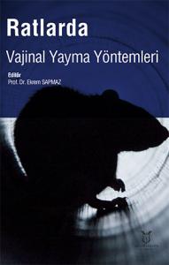
Ratlarda Vajinal Yayma Yöntemleri
Özet
Bu eser, bilimsel çalışmalarda yaygın olarak kullanılan dişi ratların üreme döngülerinin (östrus siklusu) belirlenmesi ve laboratuvar uygulamaları üzerine kapsamlı bir rehber sunmaktadır. Kitapta dişi rat genital sistem anatomisi, cinsiyet tayini kriterleri ve üreme döngüsünün hormonal mekanizmaları detaylıca açıklanmaktadır. Östrus siklusunun proöstrus, östrus, metöstrus ve diöstrus olmak üzere dört ana evresi; vajina, uterus ve overlerdeki histolojik değişiklikler ile plazma hormon konsantrasyonlarındaki dalgalanmalar üzerinden incelenmektedir. Döngü evrelerinin tayininde kullanılan invaziv olmayan makroskobik görsel değerlendirme yöntemi ile vajinal lavaj ve sürüntü alma protokolleri adım adım aktarılmaktadır. Vajinal sitolojide sıkça tercih edilen Hematoksilen & Eozin, Metilen Mavisi, Kristal Viyole, Toluidin Mavisi, Papanicolaou, Wright-Giemsa, Shorr ve Diff-Quick gibi farklı boyama tekniklerinin reaktifleri, uygulama prosedürleri ve mikroskobik sonuçları karşılaştırmalı olarak sunulmaktadır. Ayrıca, preparatlarda görülen çekirdekli epitel hücreleri, kornifiye skuamöz hücreler ve nötrofillerin oranlarına göre evre sınıflandırılması yapılarak, boyamasız direkt mikroskobik incelemenin pratik avantajlarına ve preanalitik varyasyonları önlemek için dikkat edilmesi gereken standart kriterlere değinilmektedir.
This work provides a comprehensive guide on laboratory applications and the determination of the reproductive cycle (estrous cycle) of female rats, which are widely used in scientific research. The book explains the anatomy of the female rat genital system, criteria for sex determination, and the hormonal mechanisms of the reproductive cycle in detail. The four main stages of the estrous cycle—proestrus, estrus, metestrus, and diestrus—are examined through histological changes in the vagina, uterus, and ovaries, as well as fluctuations in plasma hormone concentrations. Non-invasive macroscopic visual assessment methods, along with step-by-step protocols for vaginal lavage and smear collection used in determining cycle stages, are described. Reagents, application procedures, and microscopic outcomes of various staining techniques frequently preferred in vaginal cytology, such as Hematoxylin & Eosin, Methylene Blue, Crystal Violet, Toluidine Blue, Papanicolaou, Wright-Giemsa, Shorr, and Diff-Quick, are presented comparatively. Furthermore, stage classification based on the proportions of nucleated epithelial cells, cornified squamous cells, and neutrophils observed in preparations is detailed, while the practical advantages of unstained direct microscopic examination and the standard criteria required to prevent preanalytical variations are addressed.
Bölümler
-
Dişi Rat Genital Sistemi
-
Dişi Rat Üreme Döngüsü
-
Vajinal Lavaj ve Yayma (Smear) Yöntemi
-
Üreme Siklusunun Makroskobik Değerlendirmesi
-
Üreme Siklusunun Belirlenmesinde Kullanılan Vajinal Sitoloji Yöntemleri
-
Vajinal Sitolojide Görülen Hücre Tipleri
-
Östrus Siklus Evrelerinin Belirlenmesi
-
Farklı Vajinal Sitoloji Tekniklerinin Karşılaştırılması
-
Vajinal Sitoloji Yöntemlerinde Dikkat Edilmesi Gereken Hususlar
Referanslar
Aguilar J, Hanks M, Shaw DJ, Else R, Watson E 2006. Importance of using guarded techniques for the preparation of endometrial cytology smears in mares. Theriogenology. 66 (2): 423-430.
Ajayi, A.F., Akhigbe, R.E. Staging of the estrous cycle and induction of estrus in experimental rodents: an update. Fertil Res and Pract 6, 5 (2020).
Andrews WW, Mizejewski GJ, Ojeda SR. Development of estradiol-positive feedback on luteinizing hormone release in the female rat: a quantitative study. Endocrinology 1981, 109(5), 1404-1413.
Auta T, Hassan AT. Alteration in oestrus cycle and implantation in Mus musculus administered aqueous wood ash extract of Azadirachta indica (neem). Asian Pacific J Reproduction. 2016;5(3):188–92.
Bancroft, G. D. & Stevens, A. Theory and Practice of Technique. 4th ed. New York, Churchill Livingstone Inc., 1996
Barret KE, Barman SM, Boitano S, Brooks HL. Reproductive development and function of the female reproductive system. In: Ganong’s review of medical physiology. 24th edition, McGraw Hill Education.
Cora, M. C., Kooistra, L., & Travlos, G. (2015). Vaginal cytology of the laboratory rat and mouse: review and criteria for the staging of the estrous cycle using stained vaginal smears. Toxicologic pathology, 43(6), 776-793.
Cowie AT. Lactation. In: Austin CR, Short RV, editors. Reproduction in Animals, Volume 3, New York: Cambridge University Press; 1984. p. 195–231.
Dluzen DE, Ramirez VD. In vivo LH-RH output of ovariectomized rats following estrogen treatment, Neuroendocrinology 1986, 43(4), 459-465.
Ekambaram G, SKS K, Joseph LD. Comparative Study on the Estimation of Estrous Cycle in Mice by Visual and Vaginal Lavage Method. J Clin Diagnostic Res. 2017;11(1):AC05–7.
Findlay JK. An update on the roles of inhibin, activin, and follistatin as local regulators of folliculogenesis. Biology of Reproduction 1993, 48(1), 15-23.
Fortune JE. Ovarian follicular growth and development in mammals. Biology of reproduction 1994, 50(2) 225-232.
Freeman ME. The Ovarian Cycle of the Rat. In: Physiology of Reproduction. Editor: E. Knobil and J. Neill. 1988, Raven Press.
Gal A, Lin PC, Barger AM, MacNeill AL, Ko C 2014. Vaginal fold histology reduces the variability introduced by vaginal exfoliative cytology in the classification of mouse estrous cycle stages. Toxicol. Pathol. 42 (8): 1212-1220
Gallo RV. Pulsatile LH release during periods of low level LH secretion in the rat estrous cycle. Biology of Reproduction 1981, 24(4), 771-777.
Hafez ESE, Lea & Febiger. Reproduction and Breeding Techniques for Laboratory Animals. 1970, Philadelphia.
Haisenleder DJ, Ortolano GA, Dalkin AC, Ellis TR, Paul SJ, Marshall JC. Differential regulation of gonadotropin subunit gene expression by gonadotropin-releasing hormone pulse amplitude in female rats. Endocrinology 1990, 127(6), 2869-2875.
Hamid HY, Zakaria MZAB. Reproductive characteristics of the female laboratory rat. African journal of Biotechnology 2013, 12(19), 2510-2514.
Harkness JE, Wagner JE. The Biology and Medicine of Rabbits and Rodents.1989, Philadelphia.
Hong H. Changes in the mouse estrus cycle in response to BRCA1 inactivation suggest a potential link between risk factors for familial and sporadic ovarian cancer. Cancer research. 2010;70:221–228.
Hubscher, C. H., Brooks, D. L., and Johnson, J. R. (2005). A quantitative method for assessing stages of the rat estrous cycle. Biotech Histochem 80, 79–87.
Krafts, K. P. & Pambuccian, S. E. Romanowsky staining in cytopathology: history, advantages and limitations. Biotech. Histochem., 86(2):82-93, 2011.
Levine JE, Norman RL, Gliessman PM, Oyama TT, Bangsberg DR, Spies HG. In vivo gonadotropin-releasing hormone release and serum luteinizing hormone measurements in ovariectomized, estrogen-treated rhesus macaques. Endocrinology 1985, 117(2), 711-721.
Levine JE, Duffy MT. Simultaneous measurement of luteinizing hormone (LH)-releasing hormone, LH, and follicle-stimulating hormone release in intact and short-term castrate rats. Endocrinology 1988, 122(5), 22112221.
Levine R, Levine S. Role of the pituitary-adrenal hormones in the acquisition of schedule-induced polydipsia. Behavioral Neuroscience 1989, 103(3), 621.
Levine JE, Bauer-Dantoin AC, Besecke LM, Conaghan LA, Legan SJ, Meredith JM, Wolfe AM. Neuroendocrine regulation of the luteinizing hormone-releasing hormone pulse generator in the rat. In Proceedings of the 1990 Laurentian Hormone Conference 1991, 97-153.
Lohmiller JJ, Swing SP. Reproduction and Breeding in The Laboratory Rat. 2006.
Marcondes, F. K., L. C. M. Vanderlei, L. L. B. Lanza, and R. C. Spadari-Bratfisch, 1996: Stress-induced subsensitivity to catecholamines depends on the estrous cycle. Can. J. Physiol. Pharmacol. 74, 663–669.
McLean, A. C., Valenzuela, N., Fai, S., & Bennett, S. A. (2012). Performing vaginal lavage, crystal violet staining, and vaginal cytological evaluation for mouse estrous cycle staging identification. JoVE (Journal of Visualized Experiments), (67), e4389.
Mendoza‐Rodríguez CA, Merchant‐Larios H, Segura‐Valdez ML, MorenoMendoza N, Cruz ME, Arteaga‐López P, Cerbón M. C‐fos and estrogen receptor gene expression pattern in the rat uterine epithelium during the estrous cycle. Molecular Reproduction and Development: Incorporating Gamete Research 2003, 64(4), 379-388.
Mohammed S, Sundaram V. Comparative Study of Metachromatic Staining Methods in Assessing the Exfoliative Cell Types During Oestrous Cycle in Sprague-Dawley Laboratory Rats. Int. J. Morphol. 2018, vol.36, n.3, pp.962968.
Monroe SE, Rebar RW, Gay VL, Midgley Jr AR. Radioimmunoassay determina tion of luteinizing hormone during the estrous cycle of the rat. Endocrinology 1969, 85(4), 720-724.
OECD. Guidance or genetic document histological evaluation of endocrine and reproductive tests in rodents. Preparation, reading and reporting of vaginal smears; 2009.
Ochei, J. & Kolhatkar, A. Medical Laboratory Science: Theory and Practice. New York, McGraw-Hill Education, 2000. pp.513-37.
Paccola, Camila C, Catarina G. Resende, Taiza Stumpp, Sandra Maria Miraglia and Ivone Martins Cipriano. “The rat estrous cycle revisited: a quantitative and qualitative analysis.” Animal reproduction 10 (2013): 677-683.
Peluso JJ. Morphologic and physiologic features of the ovary. Pathobiology of The Aging Rat 1992, 1, 337-349.
Piacsek BE, Schneider TC, Gay VL. Sequential study of luteinizing hormone (LH) and “progestin” secretion on the afternoon of proestrus in the rat. Endocrinology 1971, 89(1), 39-45.
Poyraz Ö. Laboratuvar Hayvanları Yetiştiriciliği. Ankara Üniv. Vet. Fak. Yayınları. 1997.
Raimondi, G. M., Eng, A. K., Kenny, M. P., Britting, M. A., & Ostroff, L. E. (2022). Track-by-Day: A standardized approach to estrous cycle monitoring in biobehavioral research. bioRxiv.
Richardson VC, Backues K. Diseases of small domestic rodents. Journal of Zoo and Wildlife Medicine 2003, 34(4), 421-421.
Russell LD. Normal development of the testis. In: Mohr U,Dungworth DL, Capen CC, editors. Pathobiology in the Aging Rat, Volume 1, Washington, D.C: ILSI Press; 1992. p. 395–405.
Sapmaz E, Ayar A, Celik H, Sapmaz T, Kilic N, Yasar MA. Effects of melatonin and oxytetracycline in autologous intraperitoneal ovary transplantation in rats. Neuro Endocrinology Letters. 2003 Oct;24(5):350-354.
Sapmaz T, Sevgin K, Topkaraoglu S, Tekayev M, Aktas S, Coskun G, Polat S, Sapmaz E, & Irkorucu O. Comparison of melatonin, oxytetracycline, and N-acetylcysteine pre-treatments in autologous intraperitoneal ovarian transplantation in rats. Biochemical and biophysical research communications, 2022, 606, 49–54.
Smith MS, Freeman ME, Neill JD. The control of progesterone secretion during the estrous cycle and early pseudopregnancy in the rat: prolactin, gonadotropin and steroid levels associated with rescue of the corpus luteum of pseudopregnancy. Endocrinology 1975, 96(1), 219-226.
Spornitz UM, Socin CD, Dravid AA. Estrous stage determination in rats by means of scanning electron microscopic images of uterine surface epithelium. The Anatomical Record: An Official Publication of the American Association of Anatomists 1999, 254(1), 116-126.
Srinivasan MR, Sabarinathan A, Geetha A, Shalini K, Sowmiya M. A Comparative Study on Staining Techniques for Vaginal Exfoliative Cytology of Rat. J of Pharmacol & Clin Res. 2017; 3(3): 555615.
Urbanski HF, Ojeda SR. The juvenile-peripubertal transition period in the female rat: establishment of a diurnal pattern of pulsatile luteinizing hormone secretion. Endocrinology 1985, 117(2), 644-649.
Yener, T.; Turkkani Tunc, A.; Aslan, H.; Aytan, H. & Cantug Caliskan, A. Determination of oestrous cycle of the rats by direct examination: how reliable? Anat. Histol. Embryol., 36(1):75-7, 2007.
Yockim JM, Shirer HW (1981). Evidence for a photoperiod-sensitive pacemarker for estrous cycle of the rat. American Journal of Physiology, 241 (3) E261-7.
Zenclussen, M. L., Casalis, P. A., Jensen, F., Woidacki, K., & Zenclussen, A. C. (2014). Hormonal fluctuations during the estrous cycle modulate heme oxygenase-1 expression in the uterus. Frontiers in endocrinology, 5, 32.
Yayınlanan
Lisans
LisansBu İnternet Sitesi içeriğinde yer alan tüm eserler (yazı, resim, görüntü, fotoğraf, video, müzik vb.) Akademisyen Kitabevine ait olup, 5846 sayılı Fikir ve Sanat Eserleri Kanunu ve 5237 sayılı Türk Ceca Kanunu kapsamında korunmaktadır. Bu hakları ihlal eden kişiler, 5846 sayılı Fikir ve Sanat eserleri Kanunu ve 5237 sayılı Türk Ceza Kanununda yer alan hukuki ve cezai yaptırımlara tabi olurlar. Yayınevi ilgili yasal yollara başvurma hakkına sahiptir.
